
An Introduction to the Cell - SimpleMed
Cells are the units that make up the human body - each cell has a role
within the body for which it is designed and specialised (e.g.
protection - keratinized squamous epithelial cells of the skin - or
secretion - mucous-secreting columnar goblet cells of the respiratory
tract). This article covers the common components of cells in the form
of the hypotheical "standard" eukaryotic cell (cells with enveloped
nuclei).
An organelle is a component of the cell with a specific function.

Diagram - The eukaryotic cell, with its various organelles labelled, each with a specific function
Creative commons source by 3.2 The Cytoplasm and Cellular Organelles by Rice University [CC BY-SA 4.0 (https://creativecommons.org/licenses/by-sa/4.0)]
- The main central organelle of the cell.
- Contains DNA, nucleoproteins (nucleic acids bonded to a protein) & RNA.
- Function: stores DNA and co-ordinate the cell’s activities, such as growth, metabolism, protein synthesis and cell division.
- Transmission electron microscope images of the nucleus reveal that DNA is found in two forms. Heterochromatin (condensed DNA (chromatin) -> APPEARS DARK- heterochromatin has no active RNA synthesis as it is condensed) and euchromatin (uncondensed DNA -> APPEARS LIGHT - has active RNA synthesis).
- N.B. Prokaryotic cells will not have a nucleus which is what defines them as prokaryotc.
- Found within the nucleus,
- Function: the site of ribosomal RNA (rRNA) synthesis. The site at which it is combined with proteins to generate incomplete ribosomes. These ribosomes then mature after being exported from the nucleus into the cytoplasm, where they either attach to rough endoplasmic reticulum or become free-floating ribosomes within the cytoplasm.
- A double layered membrane bounding the nucleus.
- Contains nuclear pores - these allow macromolecules to pass through when entering or exiting the nucleus.
- Function: controls what enters and exits the nucleus.
Rough Endoplasmic Reticulum (RER)
- A series of interconnecting membranes, vesicles & cisternae (flattended sacs), continuous throughout the cytoplasm.
- Ribosomes are attached to the outer surface of the membranes, making this ER "rough".
- Function: synthesises proteins destined for lysosomes, the cell membrane or extracellular export.
- Pancreatic cells (and other cells which synthesise substances) have abundant RER to synthesise pancreatic enzymes.
Smooth Endoplasmic Reticulum (SER)
- A series of interconnecting membranes, vesicles and cisternae (less flat compared to RER) that are less extensive throughout the cytoplasm. The lack of embedded ribosomes classifies this ER as smooth.
- Function: lipid biosynthesis and intracellular transport (e.g. steroid production).
- Composed of two rRNA subunits (40s+60s for eukaryotes and 50s +30s for prokaryotes) that wrap around mRNA to begin translation and protein synthesis.
- Function: site of protein synthesis (translation) within the cell.
- Semi-circular shaped stacks of cisternae.
- Vesicles containing proteins bud off from the RER and fuse with the convex forming face of Golgi body.
- The Golgi bodies have polarity - proteins move from the convex (entry point) to concave end of the stack and are modified as they move.
- Function: sort, concentrate, package and modify proteins synthesised in the RER.
- Vesicles containing different proteins leave the maturing, concave face of the Golgi body, destined for lysosome assembly - they then undergo further processing or secretion.
- Membrane bound organelles that contains acid hydrolases at pH 5 (proteases, nucleases, glycosidases, lipases and phosphatases).
- Function: break down excess or worn out organelles and digested viruses or bacteria.
- A membrane bound organelle that contains enzymes that transfer hydrogen atoms from toxins such as alcohol to oxygen to produce hydrogen peroxide.
- The toxins ingested are neutralised and the hydrogen peroxide produced can be neutralised by the enzymes in peroxisomes to produce water and oxygen.
- Function: chemical detoxification and lipid metabolism.

Diagram - The structure of the mitochondria
Creative commons source by Mariana Ruiz Villarreal LadyofHats [CC BY-SA 4.0 (https://creativecommons.org/licenses/by-sa/4.0)]
- A key component of the cell. Particularly abundant in highly active, energy requiring cells.
-
Mitochondria have a double membrane with an outer and inner lipid
bilayer membrane.
- The inner membrane is larger than the outer membrane and is therefore highly folded to fit within the outer membrane. This folding increases the surface area for ATP generation to occur - these folds are called cristae.
- Contained within the inner membrane lies the matrix where the TCA/Krebs cycle takes place. The matrix contains mitochondrial DNA, ribosomes and soluble enzymes.
- The enzymes in the matrix are responsible for the creation of ATP in the TCA/Krebs cycle and oxidative phosphorylation.
- Cells such as muscle cells and sperm cells have a higher number of mitochondria due to increased requirements for energy in the form of ATP.
-
Function: Responsible for the generation of ATP viz. energy, for the cell
through aerobic respiration and regulates cellular
metabolism

Diagram - Labelled image of a mitochondria under a microscope
Creative commons source by CNX OpenStax [CC BY-SA 4.0 (https://creativecommons.org/licenses/by-sa/4.0)]
- The plasma membrane is composed of phospholipids. These are essentially triacyl glycerides (TAGs) with a phosphate head instead of a fatty acid chain. Phospholipids are amphipathic molecules (have both hydrophobic and hydrophilic regions)
- There are proteins embedded in the plasma membrane. These can be either peripheral proteins (only exist on one side of the membrane) or integral proteins (span the width of the bilayer).
- Cholesterol is present in plasma membranes and controls the fluidity or rigidity of the membrane at various temperatures.
- Take a look at our Biological membranes article for more detail on structure of membranes
-
Functions:
- Act as a selectively permeable barrier as it only allows non-polar, small molecules through without the aid of channels.
- Compartmentalises cells into discreet units.
- Communication with other molecules and cells.
- Recognition through signalling molecules, adhesion proteins and immune surveillance.
- Electrical or chemical signal generation in response to a stimuli.
- All cells posses a cytoskeleton which is responsible for maintaining and changing the cell’s shape.
- There are three main structural components of the cytoskeleton: microfilaments, intermediate filaments and microtubules.
-
Function:
- Provides structural support for the plasma membrane & cell organelles.
- Provides a means of movement for organelles, plasma membrane and other cytosol constituents around the cell.
- Provides the locomotor mechanism for amoebic movement of cells such as lymphocytes (crawling-like movement) and is the basis of cilia and flagella.
- Provides contractility in cells of specialised tissues (e.g. muscle cells).
-
Microfilaments:
- Composed of two strings of actin and measure 5nm in diameter.
- They are associated with ATP allowing for contractility. They can also assemble and dissociate very quickly (dynamic).
- The structure of microvilli of the intestinal cells is maintained by a core of actin filaments.
-
Intermediate filaments:
- Unlike microfilaments, they are not dynamic and are 10-12nm in diameter.
- Their function is to form a tough supporting meshwork within the cytoplasm. They are anchored to the plasma membrane at desmosomes (strong intracellular junctions).
- The filaments are commonly found in nerve cells, neuroglial cells and epithelial cells (made out of cytokeratin).
-
Microtubules:
- Tubulin subunits polymerise to form the wall of the hollow microtubule.
- They originate from the centrosome and found at sites in the cell where structures are moved - nerve fibres, mitotic spindles, cores of cilia & flagella.
- Attachment proteins can bind to the organelles and move the structures along the microtubules e.g. vesicles moving through the cytoplasm to the plasma membrane.
Edited by: Ben Appleby and Marcus Judge
https://simplemed.co.uk/subjects/cell-physiology-and-biology/introduction-to-the-cell
Cell Organelles- Structure and Functions with labeled diagram - by Anupama Sapkota & Microbe Notes
Cell Organelles definition
- Cell organelle is a specialized entity present inside a particular type of cell that performs a specific function.
- There are various cell organelles, out if which, some are common in most types of cells like cell membranes, nucleus, and cytoplasm. However, some organelles are specific to one particular type of cell-like plastids and cell walls in plant cells.
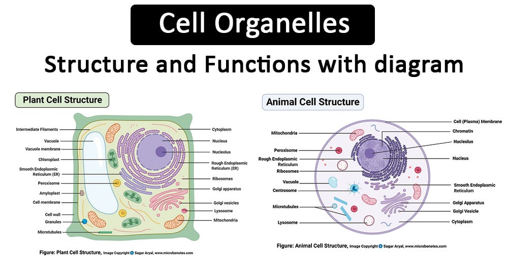
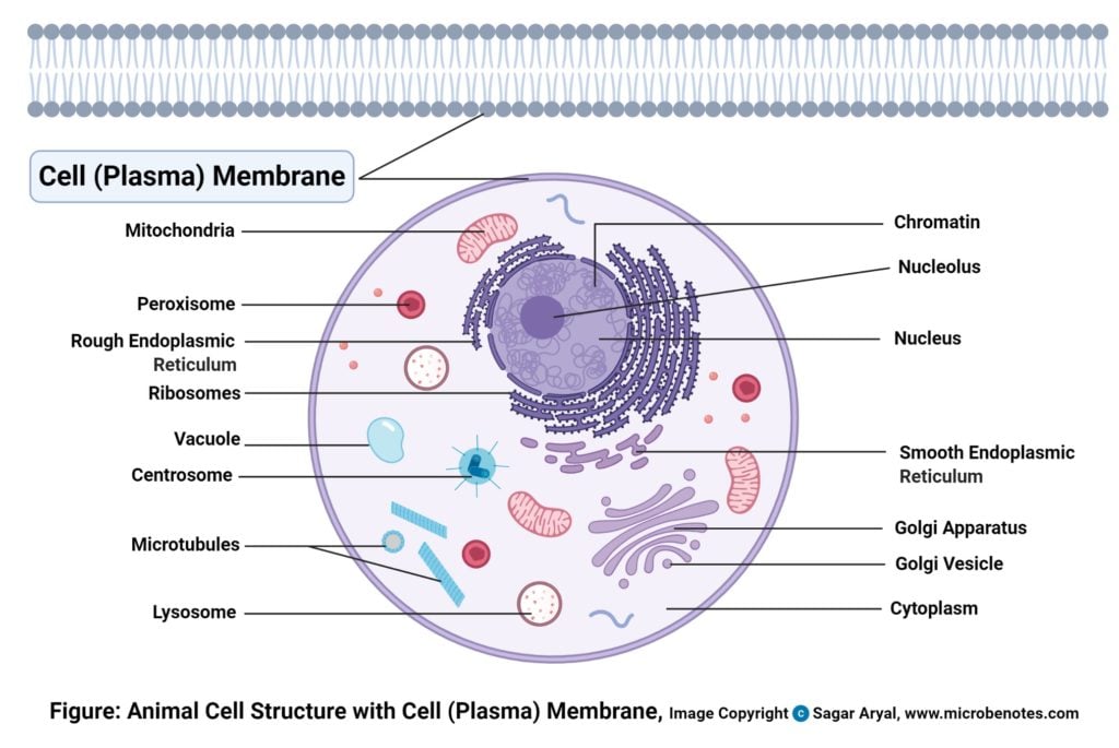
Cell membrane (Plasma membrane/ Plasmalemma)
- A plasma membrane is composed of lipids and proteins where the composition might fluctuate based on fluidity, external environment, and the different stages of development of the cell.
Structure
- Structurally, it consists of a phospholipid bilayer along with two types of proteins viz. embedded proteins and peripheral proteins that function in providing shape and allowing the movement of particles in and out of the cell.
- The most abundant lipid which is present in the cell membrane is a phospholipid which contains a polar head group attached to two hydrophobic fatty acid tails.
- The embedded proteins act as channels for the transfer of particles across the cell with some proteins acting as receptors for the binding of various components.
- The peripheral proteins function as to provide fluidity as well as mechanical support to the structure of the cell.
Functions
- The cell membrane provides mechanical support that facilities the shape of the cell while enclosing the cell and its components from the external environment.
- It regulates what can be allowed to enter and exit the cell through channels, acting as a semi-permeable membrane, which facilities the exchange of essential compounds required for the survival of the cell.
- It generates and distributes signals in and outside of the cell for the proper functioning of the cell and all the organelles.
- It allows the interaction between cells required during tissue formation and cell fusion.
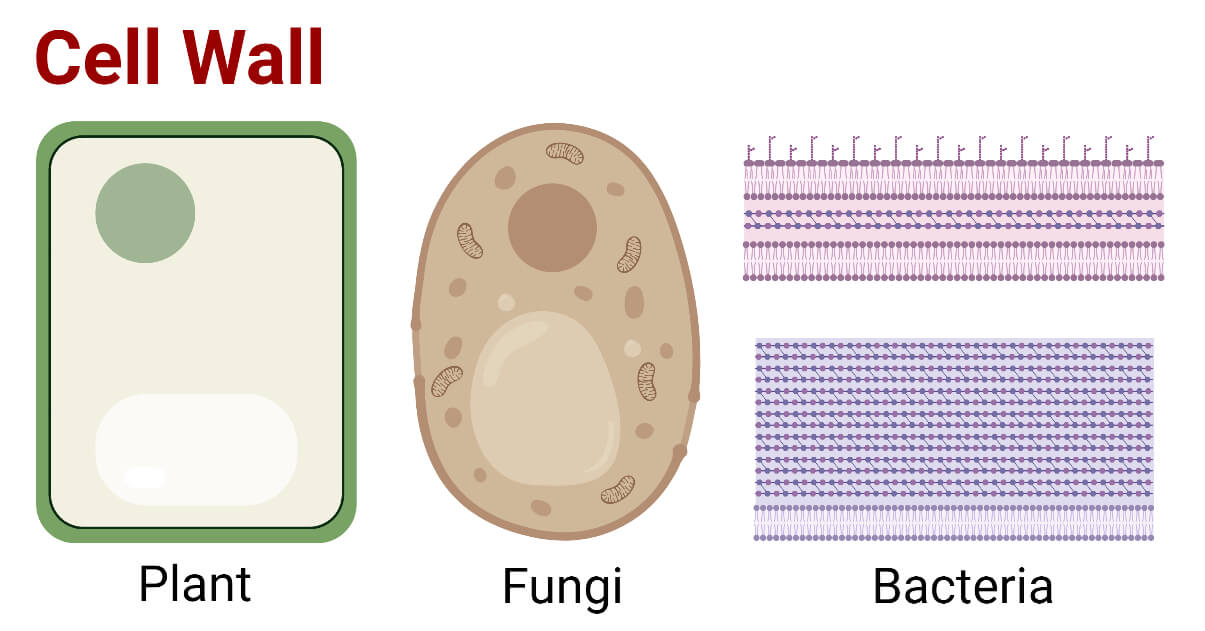
Cell Wall
- An additional non-living layer present outside the cell membrane in some cells that provides structure, protection, and filtering mechanism to the cell is the cell wall.
Structure
- In a plant cell, the cell wall is made up of cellulose, hemicellulose, and proteins while in a fungal cell, it is composed of chitin.
- A cell wall is multilayered with a middle lamina, a primary cell wall, and a secondary cell wall.
- Middle lamina contains polysaccharides that provide adhesion and allows binding of the cells to one another.
- After middle lamina is the primary cell wall which is composed of cellulose. The last layer, which is not always present, is the secondary cell wall made of cellulose and hemicellulose.
Functions
- The critical function of the cell wall is protecting and maintaining the shape of the cell. It also helps the cell withstand the turgor pressure of the cell.
- It initiates cell division by providing signals to the cell and allows the passage of some molecules into the cell while blocking others.
Centriole
- Centrioles are tubular structures mostly found in eukaryotic cells which are composed mainly of the protein tubulin.
Structure
- A centriole consists of a cylindrical structure made with nine triplets microtubules that surround the periphery of the centriole while the center has a Y-shaped linker and a barrel-like structure that stabilizes the centriole.
- Another structure called cartwheel is present in a centriole which is made up of a central hub with nine spokes/filaments radiating from it. Each of these filaments/spokes is connected to the microtubules through a pinhead.

Functions
- During cell division, centrioles have a crucial role in forming spindle fibers which assist the movement of chromatids towards their respective sides.
- They are involved in the formation of cilia and flagella.
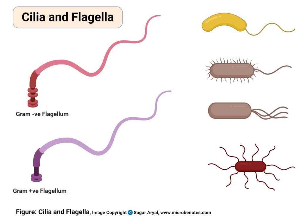
Cilia and Flagella
- Cilia and Flagella are tiny hair-like projections from the cell made of microtubules and covered by the plasma membrane.
Structure
- Cilia are hair-like projections that have a 9+2 arrangement of microtubules with a radial pattern of 9 outer microtubule doublet that surrounds two singlet microtubules. This arrangement is attached to the bottom with a basal body.
- Flagella is a filamentous organelle, the structure of which, is different in prokaryotes and eukaryotes.
- In prokaryotes, it is made up of the protein called flagellin wrapped around in a helical manner creating a hollow structure at the center throughout the length.
- In eukaryotes, however, the protein is absent and the structure is replaced with microtubules.
Functions
- The most critical role of cilia and flagella is movement. These are responsible for the movement of the organisms as well as for the movement of various particles present around the organisms.
- Some cilia present in some particular organs may have the function of sense. The cilium in the blood vessels, which helps in controlling the flow of blood is an example.
Chloroplast
- A chloroplast is a type of plastic that is involved in photosynthesis in plants and algae.
- Chloroplast contains an essential pigment called chlorophyll necessary to trap sunlight for the production of glucose.
Structure
- It is a double-membraned structure with its own DNA which is inherited from the previous chloroplast.
- These are usually lens-shaped with shape and number varying according to cells. They have an outer membrane, an inner membrane, and a thylakoid membrane that enclosed the gel-like matric called the stroma.
- The outer and inner membrane is porous and allows transport of materials while the stroma contains DNA, chloroplast ribosomes, proteins, and starch granules.
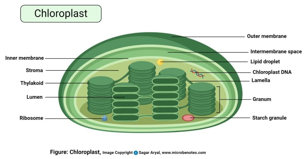
Functions
- The chloroplast is the primary center for light-dependent and light-independent reactions during photosynthesis.
- Different proteins present in chlorophyll are involved in the regulation of photorespiration.
Cytoplasm
- Cytoplasm refers to everything present inside the cell except the nucleus.
Structure
- The cytoplasm consists of a cytosol; a gel-like substance that contains other matter; cell organelles; smaller cell-like bodies bound by separate membranes; and cytoplasmic inclusions; insoluble molecules that store energy and are not surrounded by any layer.
- The cytoplasm is colorless and has about 80% water along with various nutrients required for the cell.
- It is known to have the properties of both viscous matters as well as elastic matter. Under its elasticity, cytoplasm helps in the movement of materials inside the cell by a process termed cytoplasmic streaming.
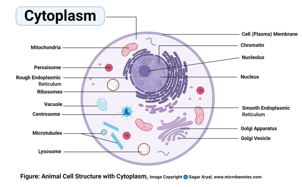
Functions
- Most of the vital cellular and enzymatic reactions like cellular respiration and translation of mRNA into proteins occur in the cytoplasm.
- It acts as a buffer and protects genetic materials as well as other organelles from damage due to collision or change in the pH of the cytosol.
- The process called cytoplasmic streaming helps in the distribution of various nutrients and facilitates the movement of cell organelles within the cell.
Cytoskeleton
- A number of fibrous structures are present in the cytosol that helps give shape to the cell while supporting cellular transport.
Structure
- Around three different classes of fibers make up the cytoskeleton which is: microtubules, microfilaments, and intermediate filaments.
- These are separated based on a protein present in them.
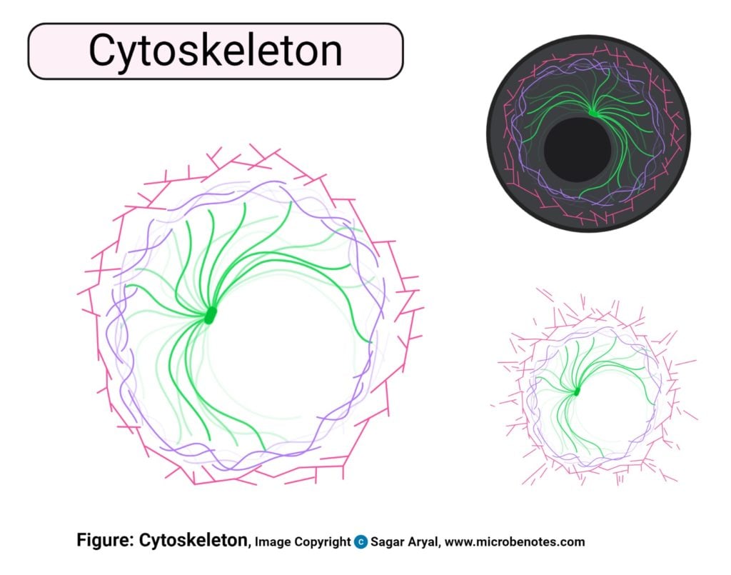
Functions
- The critical function of the cytoskeleton is to provide shape and mechanical support to the cell against deformation.
- It allows the expansion and contraction of the cell which assists in the movement of the cell.
- It is also involved in intracellular and extracellular transport of materials.
Endoplasmic Reticulum (ER)
- Endoplasmic Reticulum (ER) is present as an interconnection of tubules that are connected to the nuclear membrane in eukaryotic cells.
-
There are two types of ER based on the presence or absence of ribosomes on
them:
- Rough ER (RER) with ribosomes attached on the cytosolic face of Endoplasmic Reticulum and thus is involved in protein synthesis
- Smooth ER (SER)which lacks ribosomes and has a function during lipid synthesis.
Structure
- Endoplasmic Reticulum exists in three forms viz. cisternae, vesicles, and tubules.
- Cisternae are sac-like flattened, unbranched structures that remain stacked one on top of another.
- Vesicles are spherical structures that carry proteins throughout the cell.
- Tubules are tubular branched structures forming a connection between cisternae and vesicles.
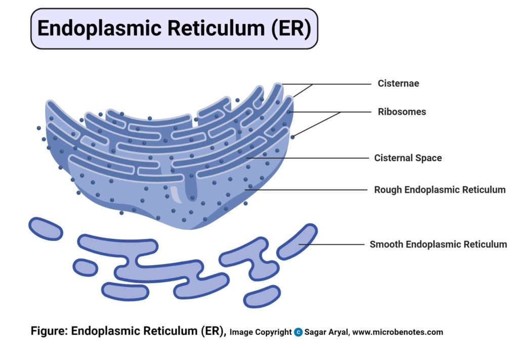
Functions
- ER contains many of the enzymes required for several metabolic processes, and the surface of the ER is essential for other operations like diffusion, osmosis, and active transport.
- One of the crucial functions of ER is the synthesis of lipids like cholesterol and steroids.
- Rough ER allows for the modification of polypeptides emerging out of the ribosomes to prepare secondary and tertiary structures of the protein.
- ER also synthesizes various membrane proteins and has a crucial role in preparing the nuclear envelope after cell division.
Endosomes
- Endosomes are membrane-bound compartments within a cell originating from the Golgi network
Structure
- There are different types of endosomes based on morphology and the time it takes for the endocytosed materials to reach them.
- The early endosomes are made with the tubular-vesicular network while the late endosomes lack tubules but contain many close-packed intraluminal vesicles. The recycling endosomes are found with microtubules and are mainly composed of tubular structures.
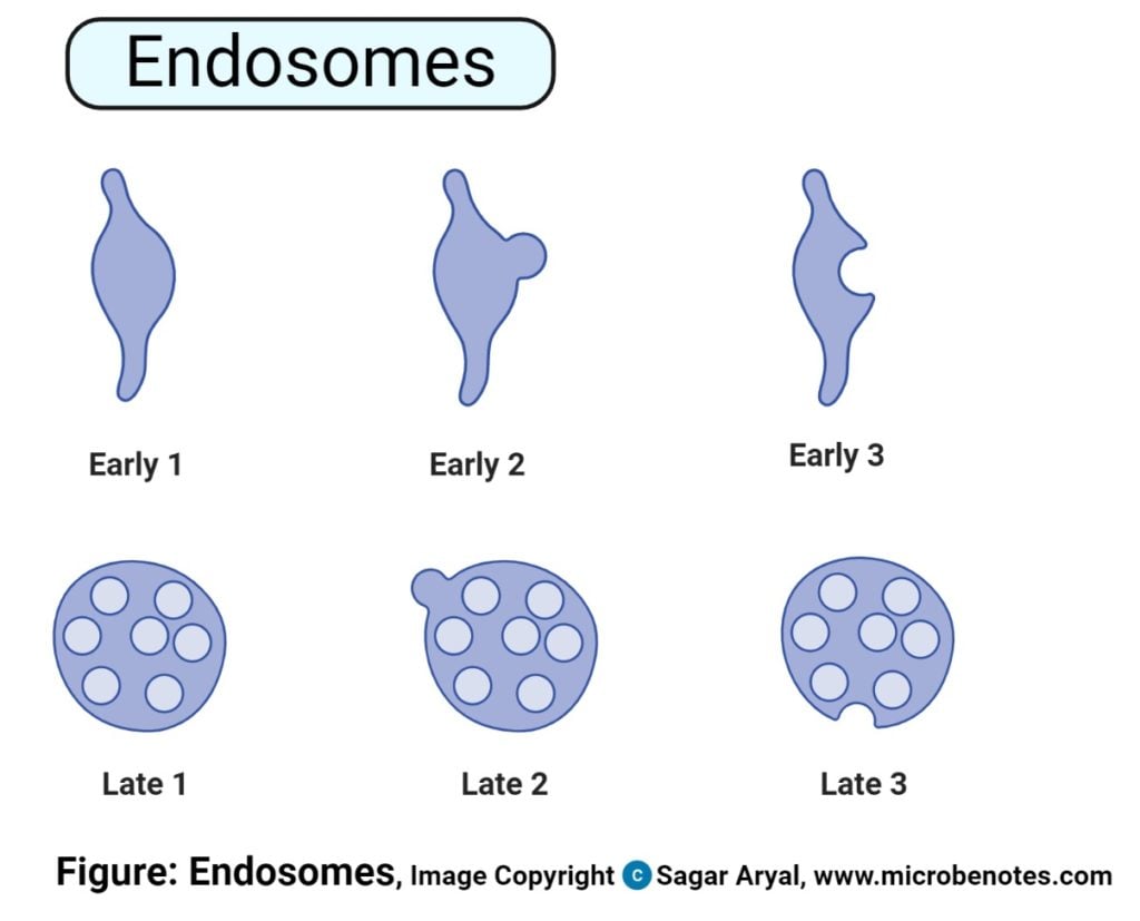
Functions
- Endosomes allow the sorting and delivery of internalized materials from the cell surface and transport of materials to the Golgi or the lysosomes.
Golgi Apparatus/ Golgi Complex/ Golgi Body
- The Golgi Apparatus is the cell organelle mostly present in eukaryotic cells which is responsible for the packaging of macromolecules into vesicles so that they can be sent out to their site of action.
Structure
- The structure of the Golgi Complex is pleomorphic; however, it typically exists in three forms, i.e. cisternae, vesicles, and tubules.
- The cisternae, which is the smallest unit of Golgi Complex, has a flattened sac-like structure which is arranged in bundles in a parallel fashion.
- Tubules are present as tubular and branched structures that radiate from the cisternae and are fenestrated at the periphery.
- Vesicles are spherical bodies that are divided into three groups as transitional vesicles, secretory vesicles, and clathrin-coated vesicles.
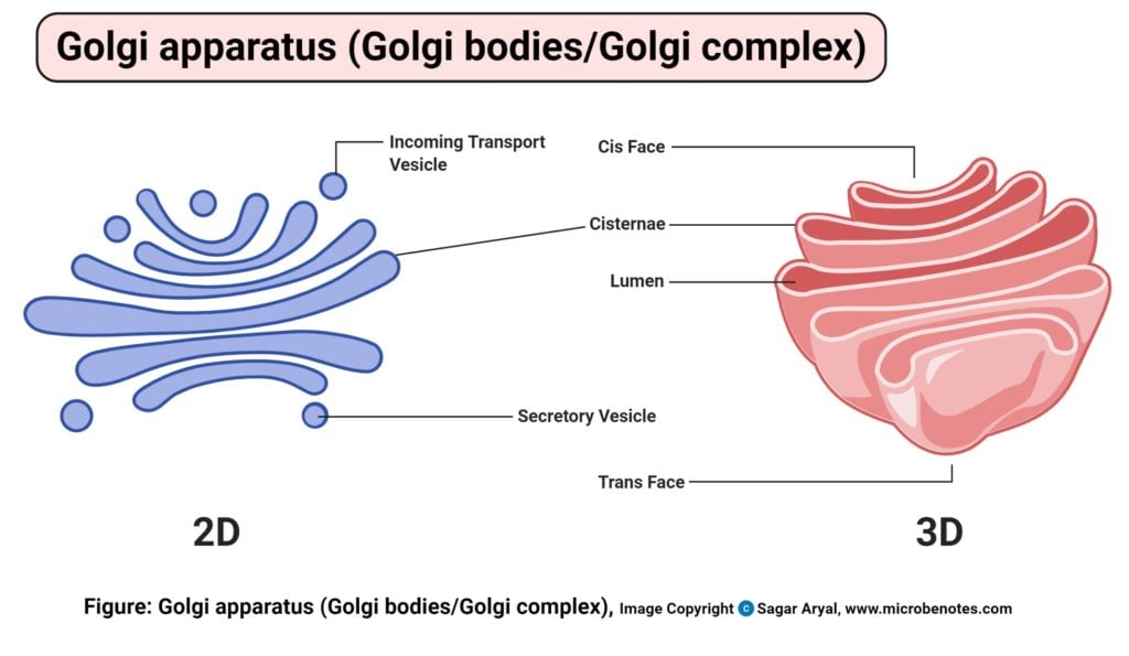
Functions
- Golgi Complex has an essential purpose of directing proteins and lipids to their destination and thus, act as the “traffic police” of the cell.
- They are involved in the exocytosis of various products and proteins like zymogen, mucus, lactoprotein, and parts of the thyroid hormone.
- Golgi Complex is involved in the synthesis of other cell organelles like a cell membrane, lysozymes, among others.
- They are also involved in the sulfation of various molecules.
Intermediate filaments
- The third class of filament that makes up the cytoskeleton are the intermediate filaments.
- They are designated at intermediate filaments because of the intermediate diameter of the filaments as compared to microfilaments and myosin proteins.
Structure
- Intermediate filaments contain a family of related proteins.
- The individual filaments are coiled around each other in a helical structure called coiled-coil structure.
Functions
- Intermediate filaments contribute to the structural integrity of a cell while playing a crucial role in holding tissues of various organs like the skin.
Lysozyme
- Lysozymes are membrane-bound organelles that occur in the cytoplasm of animal cells.
- These organelles contain an array of hydrolytic enzymes required for the degradation of various macromolecules.
-
There are two types of lysozymes:
- Primary lysosome containing hydrolytic enzymes like lipases, amylases, proteases, and nucleases.
- Secondary lysozyme formed by the fusion of primary lysozymes containing engulfed molecules or organelles.
Structure
- The shape of lysozymes is irregular or pleomorphic; however, mostly, they are found in the spherical or granular structure.
- Lysozymes are surrounded by a lysosomal membrane that contains the enzymes within the lysosome and protects the cytosol with the rest of the cell from the harmful action of the enzymes.

Functions
- These organelles are responsible for intracellular digestion where the larger macromolecules are degraded into smaller molecules with the help of enzymes present in them.
- Lysozymes also perform the critical function of the autolysis of unwanted organelles within the cytoplasm.
- Besides these, the lysosome is involved in various cellular processes, including secretion, plasma membrane repair, cell signaling, and energy metabolism.
Microfilaments
- Microfilaments are a part of the cytoskeleton of a cell made up of actin protein in the form of parallel polymers.
- These are the smallest filaments of the cytoskeleton with high rigidity and flexibility, providing strength and movement to the cell.
Structure
- The filaments are present either in cross-linked forming networks or as bundles. The chains of protein remain twisted around each other in a helical arrangement.
- One of the polar ends of the filament is positively charged and barbed, whereas the other end is negatively charged and pointed.
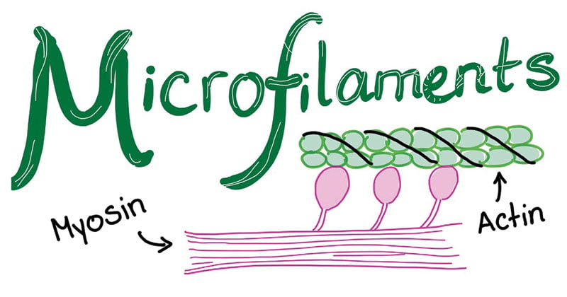
Functions
- It generates the strength for the structure and movement of the cell in association with myosin protein.
- They help in cell division and are involved in the products of various cell surface projections.
Microtubules
- Microtubules are also a part of the cytoskeleton differing from microfilaments in the presence of tubulin protein
Structure
- They are long hollow, beaded tubular structure of diameter of about 24nm.
- The wall of the microtubules consists of globular subunits present at a helical array of a and b tubulin.
- Similar to microfilaments, the ends of microtubules also have a defined polarity with one end being positively charged while the other being negatively charged.
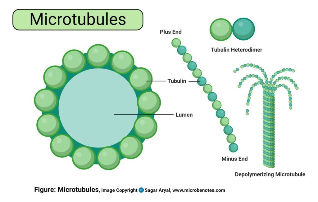
Functions
- As a part of the cytoskeleton, they provide shape and movement to the cell.
- Microtubules facilitate the movement of other cell organelles within the cell through binding proteins.
Microvilli
- Microvilli are tiny finger-like structures that project on or out of the cells. These exist either on their own or in conjunction with villi.
Structure
- Microvilli are bundles of protuberances loosely arranged on the surface of the cell with little or no cellular organelles.
- These are surrounded by a plasma membrane enclosing cytoplasm and microfilaments.
- These are bundles of actin filaments bound by fimbrin, villin, and epsin.
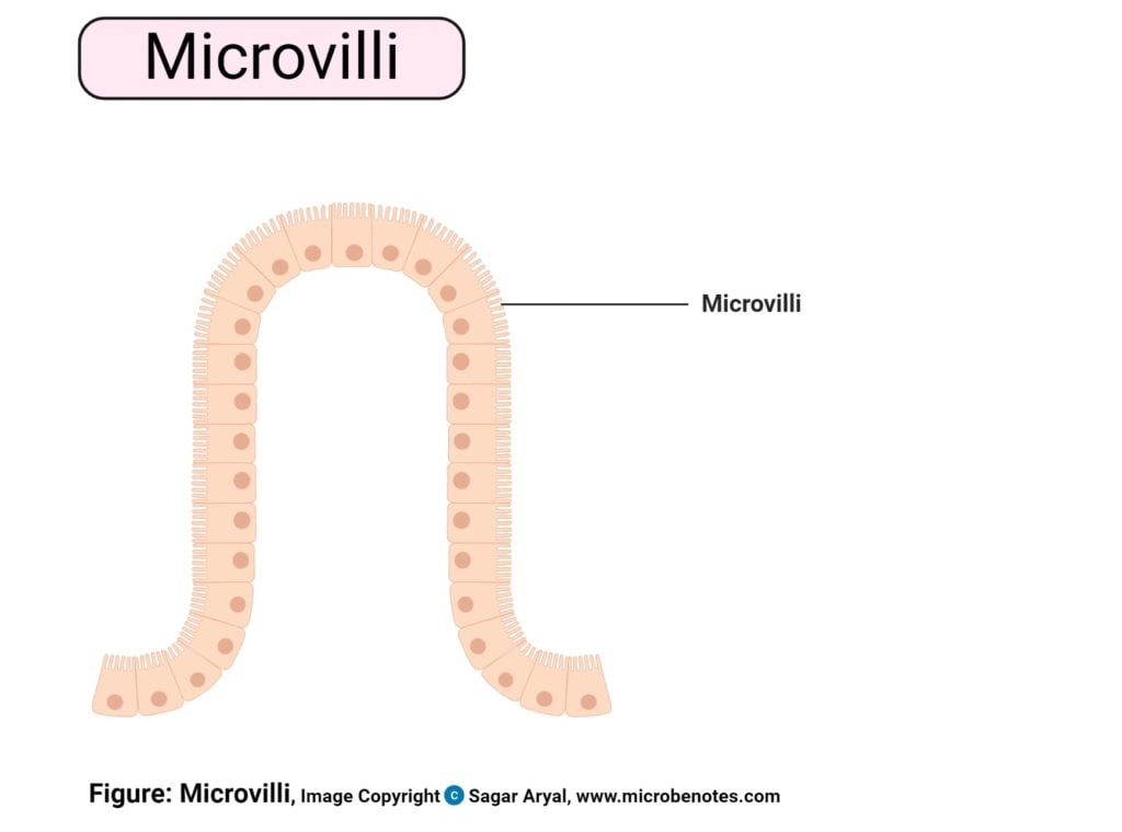
Functions
- Microvilli increase the surface area of the cell, thus, enhancing the absorption and secretion functions.
- The membrane of microvilli is packed with enzymes that allow the break down of larger molecules into smaller allowing more effective absorption.
- Microvilli act as an anchoring agent in white blood cells and in sperms during fertilization.
Mitochondria
- Mitochondria are double membrane-bound cell organelles responsible for the supply and storage of energy for the cell.
- The oxidation of various substrates in the cell to release energy in the form of ATP (Adenosine Triphosphate) is the primary purpose of mitochondria.
Structure
- A mitochondrion contains two membranes with the outer layer being smooth while the inner layer is marked with folding and finger-like structures called cristae.
- The inner mitochondrial membrane contains various enzymes, coenzymes, and components of multiple cycles along with pores for the transport of substrates, ATP, and phosphate molecules.
- Within the membranes is a matrix that contains various enzymes of metabolic processes like Kreb’s cycle.
- In addition to these enzymes, mitochondria are also home to single or double-stranded DNA called mtDNA that is capable of producing 10% of the proteins present in the mitochondria.
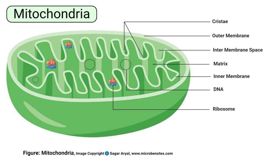
Functions
- The primary function of mitochondria is the synthesis of energy in the form of ATP required for the proper functioning of all the cell organelles.
- Mitochondria also help in balancing the amount of Ca+ ions within the cell and assists the process of apoptosis.
- Different segments of hormones and components of blood are built within mitochondria.
- Mitochondria in the liver have the ability to detoxify ammonia.
Nucleus
- The nucleus is a double membrane-bound structure responsible for controlling all cellular activities as well as a center for genetic materials, and it’s transferring.
- It is one of the large cell organelles occupying 10% of total space in the cell.
- It is often termed the “brain of the cell” as it provides commands for the proper functioning of other cell organelles.
- A nucleus is clearly defined in the case of a eukaryotic cell; however, it is absent in prokaryotic organisms with the genetic material distributed in the cytoplasm.
Structure
- Structurally, the nucleus consists of a nuclear envelope, chromatin, and nucleolus.
- The nuclear envelope is similar to the cell membrane in structure and composition. It has pores that allow the movement of proteins and RNA in and outside the nucleus. It enables the interaction with other cell organelles while keeping nucleoplasm and chromatin within the envelope.
- The chromatin in the nucleus contains RNA or DNA along with nuclear proteins, as genetic material that is responsible for carrying the genetic information from one generation to another. It is present in a sense and compact structure which might be visible as chromosome under powerful magnification.
- The nucleolus is like a nucleus within the nucleus. It is a membrane-less organelle that is responsible for the synthesis of rRNA and assembly of ribosomes required for protein synthesis.
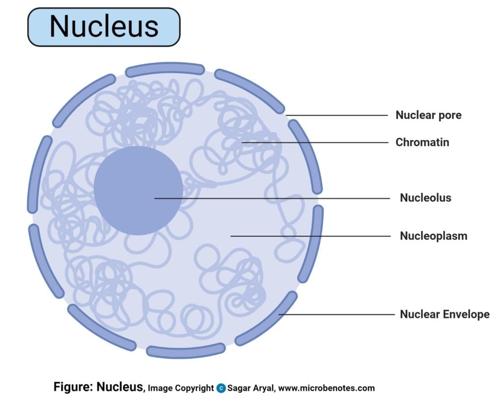
Functions
- The nucleus is responsible for storage as well as the transfer of genetic materials in the form of DNA or RNA.
- It aids in the process of transcription by the synthesis of mRNA molecules.
- The nucleus controls the activity of all other organelles while facilitating processes like cell growth, cell division and synthesis of proteins.
Peroxisomes
- Peroxisomes are oxidative membrane-bound organelles found in the cytoplasm of all eukaryotes.
- The name is accredited due to their hydrogen peroxide generating and removing activities.
Structure
- Peroxisome consists of a single membrane and granular matrix scattered in the cytoplasm.
- They exist either in the form of interconnected tubules or as individual peroxisomes.
- The compartments within every peroxisome allow the creation of optimized conditions for different metabolic activities.
- They consist of several types of enzymes with major groups being urate oxidase, D-amino acid oxidase, and catalase.
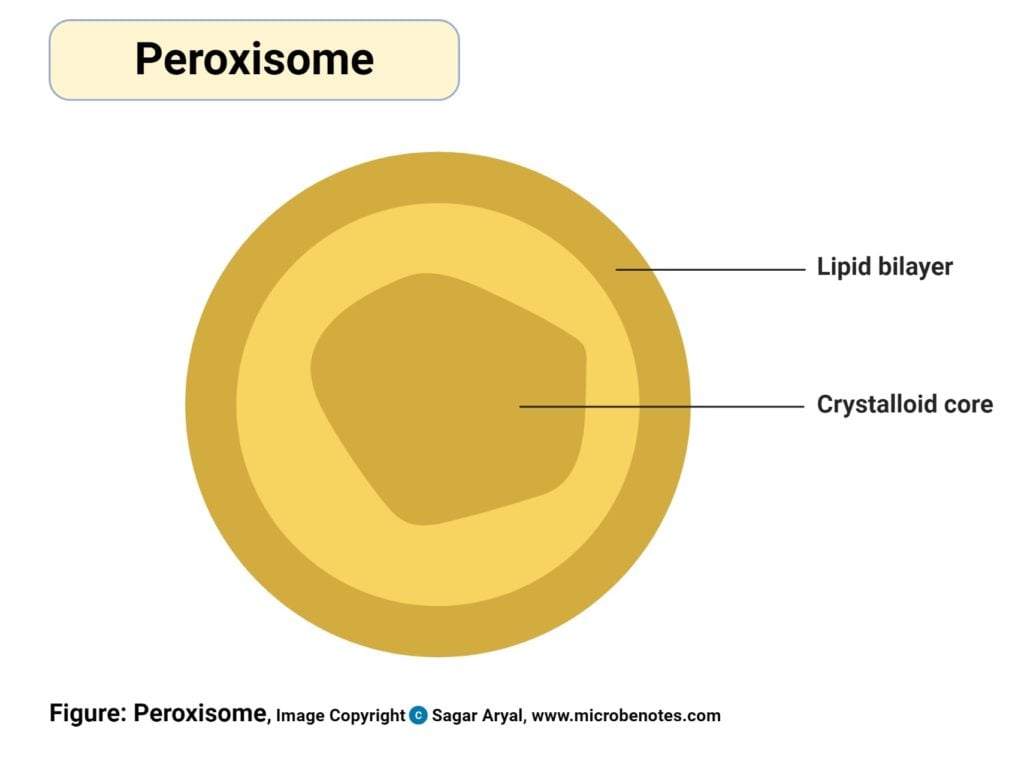
Functions
- Peroxisomes are involved in the production and elimination of hydrogen peroxide during biochemical processes.
- Oxidation of fatty acids takes place within peroxisomes.
- Additionally, peroxisomes are also involved in the synthesis of lipid-like cholesterol and plasmalogens.
Plasmodesmata
- Plasmodesmata are tiny passages or channels that allow the transfer of material and communication between different cells.
Structure
- There are 103 – 105 number of plasmodesmata connecting two adjacent cells with 50-60 nm in diameter.
-
A plasmodesma has three layers:
- The plasma membrane is continuous with the plasma membrane of the cell and has the same phospholipid bilayer.
- The cytoplasmic sleeve that is continuous with the cytosol that allows the exchange of materials between two cells.
- Desmotubule which is a part of the endoplasmic reticulum that provides a network between two cells and allows the transport of some molecules.
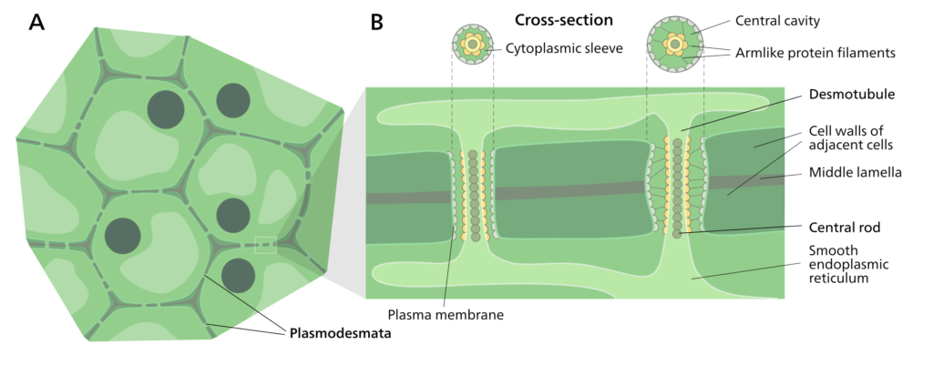
Figure: Diagram of Plasmodesmata. Source: Wikipedia
Functions
- Plasmodesmata are the primary site for the communication of two cells. It allows the transfer of molecules like proteins, RNA, and viral genomes.
Plastids
- Plastids are double membrane-bound structures present in plants and other eukaryotes involved in the synthesis and storage of food.
Structure
- Plastids are usually oval or spherical with an outer and an inner membrane between which lies the intermembrane space.
- The inner membrane enclosed a matrix called stroma that contains small structures called grana.
- Each granum consists of several sac-like thylakoids piled one on the other and connected by stroma lamellae.
- Plastids contain DNA and RNA that allows it to synthesize necessary proteins for different processes.
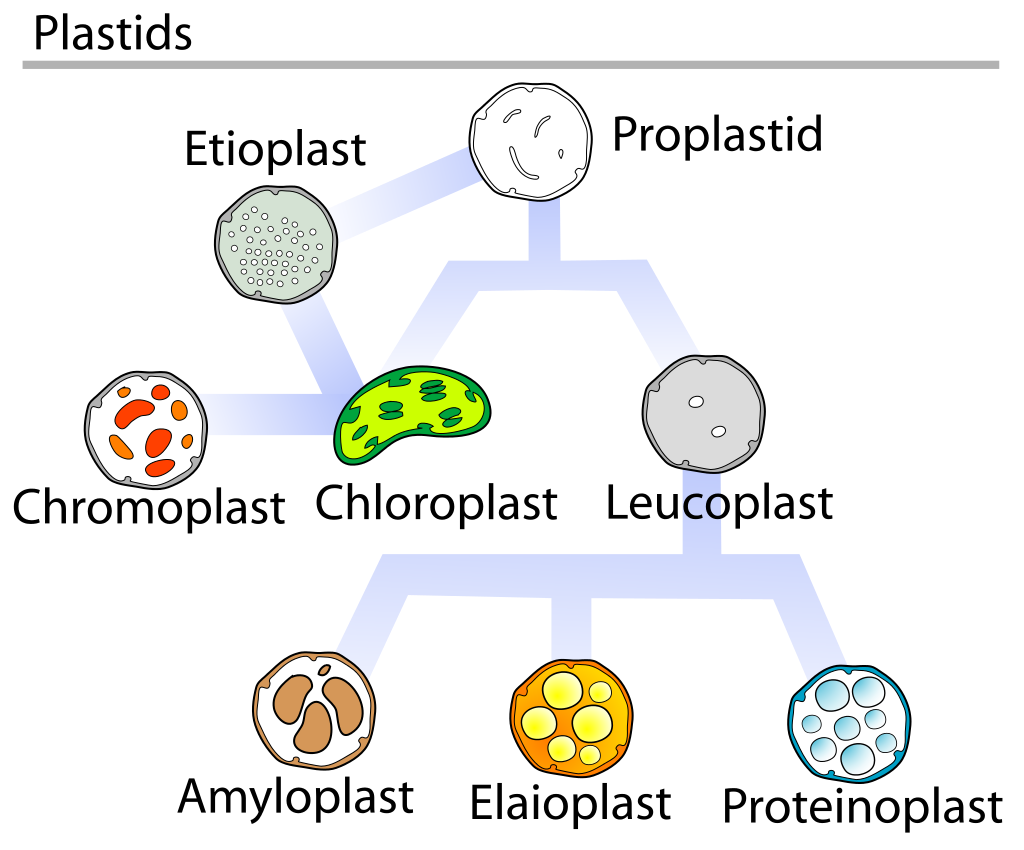
Figure: Diagram of types of plastids. Source: Wikipedia
Functions
- Chloroplasts are the center for many metabolic activities, including photosynthesis as it contains enzymes and other components required for it.
- They are also involved in the storage of food, primarily starch.
Ribosomes
- Ribosomes are ribonucleoprotein containing equal parts RNA and proteins along with an array of other essential components required for protein synthesis.
- In prokaryotes, they exist freely while in eukaryotes, they are found either free or attached to the endoplasmic reticulum.
Structure
- The ribonucleoprotein consists of two subunits.
- In the case of prokaryotic cells, the ribosomes are of the 70S with the larger subunit of 50S and the smaller one of 30S.
- Eukaryotic cells have 80S ribosomes with 60S larger subunit and 40S smaller subunit.
- Ribosomes are short-lived as after the protein synthesis, the subunits split up and can be either reused or remain broken up.
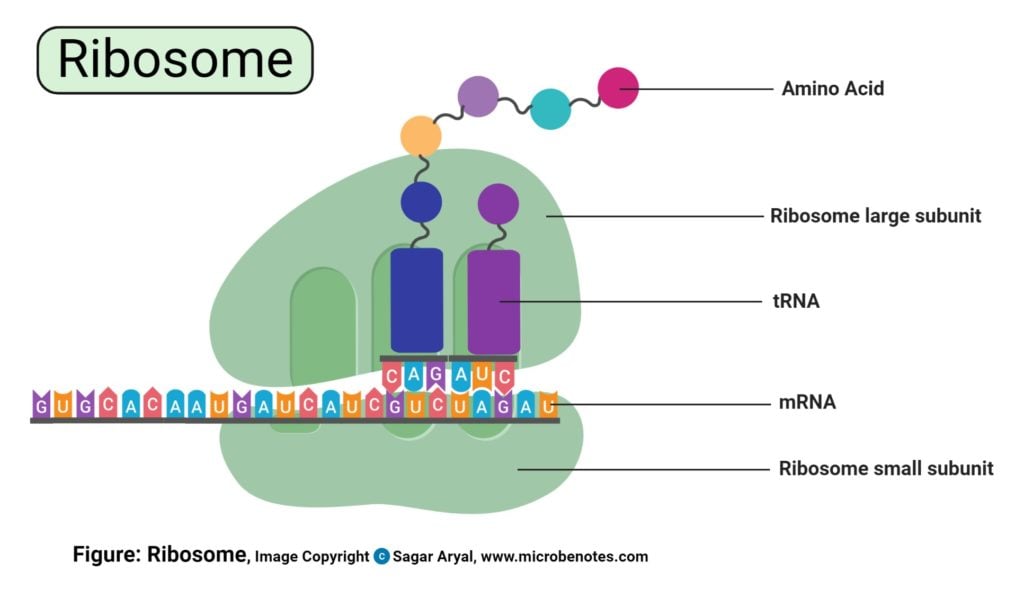
Functions
- Ribosomes are the site of biological protein synthesis in all living organisms.
- They arrange the amino acids in the order indicated by tRNA and assist in protein synthesis.
Storage granules
- Storage granules are membrane-bound organelles, also called zymogen granules storing cell’s energy reserve and other metabolites.
Structure
- These granules are surrounded by a lipid bilayer and are composed mostly of phosphorus and oxygen.
- The components inside these storage granules depend on their location in the body with some even containing degradative enzymes yet to participate in digestive activities.
Figure: Diagram of Storage Granules. Image Source: Slide Player
Functions
- Many prokaryotes and eukaryotes store nutrients and reserves in the form of storage granules in the cytoplasm.
- Sulfur granules are characteristic of prokaryotes that utilize hydrogen sulfide as a source of energy.
Vacuole
- Vacuoles are membrane-bound structures varying in size in cells of different organisms.
Structure
- The vacuole is surrounded by a membrane called tonoplast, which encloses fluid containing inorganic materials like water and organic materials like nutrients and even enzymes.
- These are formed by the fusion of various vesicles, so vacuoles are very similar to vesicles in structure.
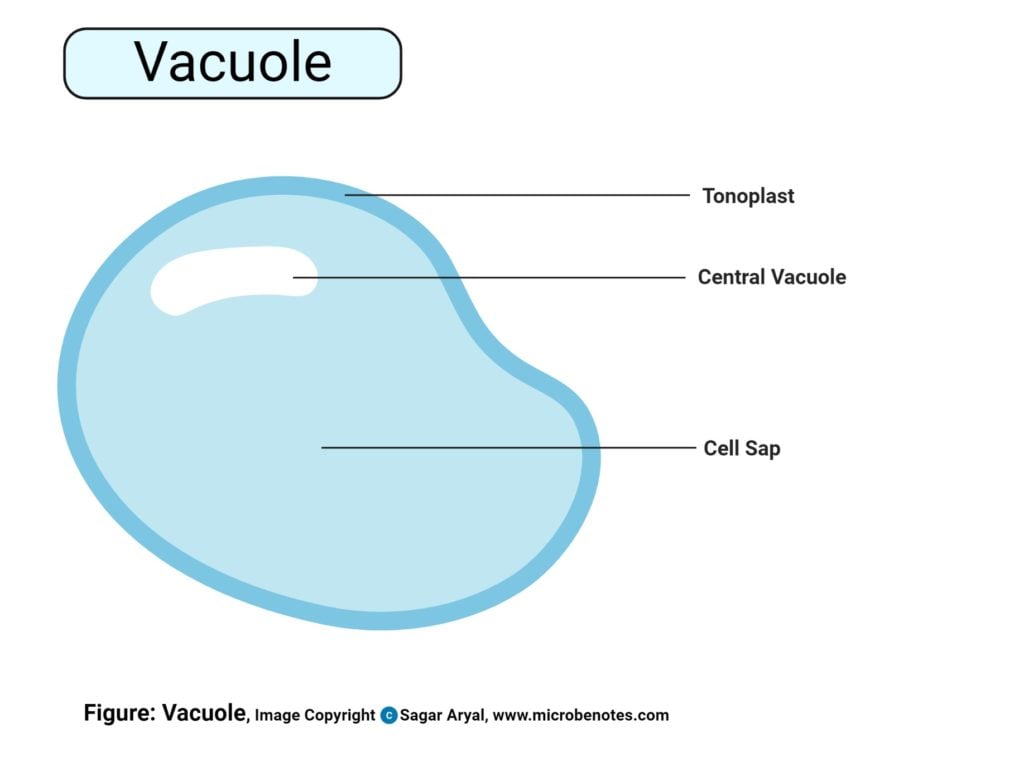
Functions
- Vacuoles act as a storage for nutrients as well as waste materials to protect the cell for toxicity.
- They have an essential function of homeostasis as it allows the balance of pH of the cell by influx and outflow of H+ ions to the cytoplasm.
- Vacuoles contain enzymes that play an important role in different metabolic processes.
Vesicles
- Vesicles are structures present inside the cell which are either formed naturally during processes like exocytosis, endocytosis or transport of materials throughout the cell, or they might form artificially, which are called liposomes.
- There are different types of vesicles like vacuoles, secretory and transport vesicles based on their function
Structure
- A vesicle is a structure containing liquid or cytosol which is enclosed by a lipid bilayer.
- The outer layer enclosing the liquid is called a lamellar phase which is similar to the plasma membrane. One end of the lipid bilayer it hydrophobic whereas the other end is hydrophilic.
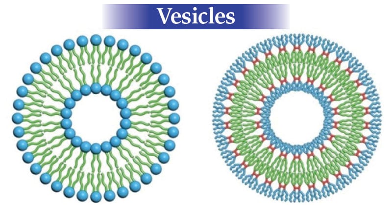
Figure: A liposome (left) and dendrimersome. The blue parts of their molecules are hydrophilic, the green parts are hydrophobic. Credit: Image courtesy of University of Pennsylvania
Functions
- Vesicles facilitate the storage and transport of materials in and outside the cell. It even allows the exchange of molecules between two cells.
- Because vesicles are enclosed inside a lipid bilayer, vesicles also function in metabolism and enzyme storage.
- They allow temporary storage of food and also control the buoyancy of the cell.
References
- https://bscb.org/learning-resources/softcell-e-learning/golgi-apparatus/
- https://micro.magnet.fsu.edu/cells/lysosomes/lysosomes.html
- https://www.britannica.com/science/mitochondrion
- https://www.khanacademy.org/science/biology/structure-of-a-cell/prokaryotic-and-eukaryotic-cells/a/plasma-membrane-and-cytoplasm
- https://www.khanacademy.org/science/biology/structure-of-a-cell/prokaryotic-and-eukaryotic-cells/a/nucleus-and-ribosomes
- https://www.ncbi.nlm.nih.gov/books/NBK9889/
- https://www.ncbi.nlm.nih.gov/books/NBK9896/
- https://www.ncbi.nlm.nih.gov/books/NBK9928/
- http://www.biology4kids.com/files/cell_vacuole.html
- http://cytochemistry.net/cell-biology/intermediate_filaments.htm
- https://www.sciencedirect.com/topics/agricultural-and-biological-sciences/plasmodesmata
- https://biologydictionary.net/vesicle/
- https://www.britannica.com/science/microvilli
- Luby-Phelps K. The physical chemistry of cytoplasm and its influence on cell function: an update. Biol. Cell. 2013;24:2593–2596.
- Lavanya, P. (2005). Cell and Molecular Biology, Rastogi Publications.
- Verma, P. S., & Agrawal, V. K. (2006). Cell Biology, Genetics, Molecular Biology, Evolution & Ecology (1 ed.). S .Chand and company Ltd.
- Images created using biorender.com
Internet Sources
Cell Organelles & Their Functions
What is cell organelle?
A cell organelle is a tiny cellular structure that performs specific functions within a cell. You can think of cell organelles as a cell’s internal organs. For example, the nucleus is the cell’s brain, and the mitochondria are the cell’s hearts. Cell organelles are often enclosed by their own membranes, which divide the cell into many small compartments for different biochemical reactions.

[In this figure] The anatomy of an animal cell with organelles labeled.
Cell organelles have a wide range of responsibilities, from generating
energy for a cell to controlling its growth and reproduction. From this
point of view, you can also think of cell organelles as different teams
within the factory. Each team carries out its specific task and
coordinates to make sure the entire factory works smoothly.
Below is a list of the cell organelles found in animal and plant cells,
which we’ll use as our guide for this discussion.
| Cell Organelle | Biological Function | Factory Part |
| Nucleus | DNA Storage | Files and blueprints management |
| Mitochondrion | Energy production | Powerplant |
| Ribosome | Protein synthesis | Machine to product toys |
| Rough ER | Protein production and modification | Coordination of toy production line and decoration |
| Smooth ER | Lipid production and Detoxification | Accessory production |
| Golgi apparatus | Protein transportation and export | Packaging and shipping department |
| Peroxisome | Lipid breakdown; redox reactions | Hazard chemical handling |
| Lysosome | Protein destruction | Recycling |
| Cytoskeleton | Cell movement; intracellular transportation | Conveyor system |
| Cell membrane | Define the inside and outside of a cell | Factory building |
| Cell wall | Structural support and protection (plant cell) | Reinforced factory building |
| Cytosol | Cellular fluid | Internal space and floor plan |
| Chloroplast | Photosynthesis (plant cell) | Solar panels |
| Vacuole | Storage and water regulation (plant cell) | Storage spaces |
Cell organelles can be divided into three types
In this article, we are going to divide these cell organelles/structures
into three types:
1. General cell organelles: they are present in both animal and plant cells all the time – cell
membrane, cytosol, cytoplasm, nucleus, mitochondrion, rough and smooth
endoplasmic reticulum, Golgi apparatus, peroxisome, lysosome, and the
cytoskeleton.
2. Temporal cell organelles: they are only found at specific stages of the cell’s life cycle –
chromosome, centrosome, autophagosome, and endosome.
3. Cell type specific cell organelles: they only exist in the plant cells – chloroplast, central vacuole, and cell wall.
Many unique cell organelles/structures only
exist in specific cell types. For example, the food vacuoles in amoeba and the trichocysts in paramecia, which cannot be found in human cells. On the other hand, some human
cells also have unique organelles that can’t be found anywhere else, like
the Weibel–Palade bodies in blood vessel cells.
1. General cell organelles in every cell
Cell membrane
- Cell membrane is a biological membrane that separates the interior of the cell from the outside space and protects the cell from its environment.
- Cell membrane is made by two layers of lipid films (oil molecules) with many kinds of membrane proteins.
- Cell membrane controls the movement of molecules such as water, ions, nutrients, and oxygen in and out of the cell.
- Proteins on the cell membrane also involved in cell movement and the communication between cells. For example, cells received signals from the outside world through different kinds of receptor proteins inserted on the cell membrane like tiny antennas.

[In this figure] The cell membrane defines the inside and outside spaces of a
cell. There are many proteins on or inserted in the cell membrane. They
function as channels (controlling the in and out of molecules) or
receptors (receiving signals from the outside world).
The image was created with
BioRender.com.
Cytosol
- Cytosol is the cellular fluid inside the cell. It fills up the entire intracellular space.
- Water is the most abundant molecule inside the cells, accounting for 70% or more of total cell mass.
- Cytosol is a complex mixture of all kinds of substances dissolved in water, including small molecules like ions (sodium, potassium, or calcium), amino acids, nucleotides (the basic DNA units), lipids, sugars, and large macromolecules such as proteins and RNA.
Cytoplasm
- Cytoplasm refers to all material within a cell, enclosed by the cell membrane, except for the cell nucleus.
- Cytoplasm includes the cytosol and all the organelles.
Cytoskeleton
- Cytoskeleton is the cells’ skeleton system. Its network reaches every inch inside the cells.
- Cytoskeleton is a dynamic network built by interlinking protein filaments. It is composed of three main components, actin filaments, intermediate filaments, and microtubules.
- Once a portion of the cytoskeleton contracts or extends, it deforms the cells and allows cells to change their shapes and movement.
- Cytoskeleton also serves as a highway system inside the cytosol. Motor proteins can carry cargos while walking along the cytoskeleton. A variety of intracellular cargoes, including proteins, RNAs, vesicles, and even entire organelles, can move around inside a cell by this intracellular transportation system.

[In this figure] Cytoskeleton consists of three types of filament proteins:
microtubules, actins, and intermediate filaments.
The image was created with
BioRender.com.

[In this figure] Fluorescent image of vimentin, an intermediate filament protein (green), in human cells. The nuclei were stained in blue color.

[In this figure] Fluorescence image of microtubule (orange), and the nucleus (cyan)
inside a cell.
Microtubule is
one type of cytoskeleton inside the cells, and it shapes cell’s
morphology. Magnification, 63x.
Photo credit: Jason Kirk, 2020 photomicrograph competition.

[In this figure] Fluorescence image of microtubule (yellow) and the nucleus (cyan)
inside a cell.
Microtubules
radiated from a tissue cell culture. Notice that the microtubules extend
to the very end of the cell membrane. Magnification, 63x.
Photo credit: Jason Kirk, 2020 photomicrograph competition.
Nucleus
- The nucleus (plural: nuclei) is a membrane-bound organelle that stores most of our genetic information (genome).
- The key feature that separates eukaryotic cells (animals, plants, and fungi) from prokaryotic cells (bacteria and archaea) is the presence of a nucleus.
- The membrane of the nucleus is called the nuclear envelope. There are nuclear pores to control transportation across the envelope.
- During cell division, the nuclear envelope will temporally disappear to allow the separation of chromosomes.
- Both DNA replication and RNA transcription happen inside the nucleus. Messager RNA (mRNA) that carries the genetic information will be exported through nuclear pores into the cytosol for protein synthesis (translation).

[In this figure] Cell nucleus is a membrane-bound organelle that stores DNA.
The image was created with BioRender.com.
Nucleolus
- Nucleolus (plural: nucleoli) is a structure inside the nucleus.
- Nucleolus is known as the site of ribosome biogenesis.
Mitochondrion
- Mitochondrion (plural: mitochondria) is a rod-shaped organelle that is considered the power generators of the cell.
- Mitochondrion performs cellular respiration, which converts glucose and oxygen to adenosine triphosphate (ATP). ATP is the biochemical energy “currency” of the cell for all activities.
- Mitochondrion has double layers of the membrane: outer mitochondrial membrane (OMM) and inner mitochondrial membrane (IMM). Between the OMM and IMM is the intermembrane space. The region inside the inner membrane is called the matrix.
- Mitochondrion generates ATP like a hydraulic dam. It happens via the electron transport chain across the IMM.
- Mitochondria (in plant cells, chloroplasts, too) are the only organelles that have their own DNA other than the nucleus. Mitochondrial DNA (mtDNA) is circular and encoded only 13 genes.
- Scientists believe mitochondria and chloroplasts are derived from the bacteria that were engulfed by the early ancestors of today’s eukaryotic cells. This theory is called the endosymbiotic theory.

[In this figure] Left: the structure of mitochondrion showing many folds of membranes and mtDNA. Right: a mitochondrion surrounded by rough ER under a transmission electron microscope.
Endoplasmic reticulum
- Endoplasmic reticulum (ER) is an internal membrane that forms branching networks of many interconnected sacs and tubes.
- There are two types of ER: rough ER and smooth ER.
- The outer side (facing the cytosol) of the rough ER is studded with ribosomes. Under the electron microscope, the dense granular ribosomes gave the name of “rough” ER.
- Rough ER stays closer to the nucleus and coordinates protein synthesis.
- Smooth ER lacks ribosomes. It specializes in lipid synthesis, steroid hormone production, and detoxification.

[In this figure] The anatomy of ER.
Left: The
relationship between the nucleus, rough, and smooth ER. Right: A 3D view
of rough ER.
The
image was created with BioRender.com.
Ribosome
- Ribosomes are the places where proteins are synthesized in our cells.
- Ribosomes consist of two major components: the small and large ribosomal subunits. They are assembled by proteins and ribosomal RNA (rRNA).
- Ribosomes translate mRNA into polypeptide chains, which fold and assemble into proteins.
- Transfer RNA (tRNA) carries the corresponding amino acid. Only the right tRNA can enter the ribosome and pair with the code on mRNA. Once the tRNA and mRNA match, the ribosome will add this amino acid onto a growing polypeptide chain.
- Ribosomes can be found on the rough ER or free-floating in the cytosol.

[In this figure] The ribosome works like a machine to translate the code sequence of mRNA into a protein.
Golgi apparatus
- Golgi apparatus (or Golgi) consists of several stacks of membrane-bound cisternae (sacs).
- Golgi apparatus usually locates close to the ER. It receives the raw protein products from the ER, modifies them (for example, adding tags made by sugar chains), and exports the proteins to a variety of destinations.
- The transportation of proteins is done within small bubbles, called vesicles.
- The vesicles are generated by budding from the membrane of the ER and Golgi. Once the vesicles reach their destinations, the fusion of membrane releases their protein cargos.
- There are three major destinations of proteins: (1) sent to other organelles, (2) released into the cytosol, and (3) secreted outside the cells. Secreting vesicles can also store the proteins until they receive a signal to release at a specific event.

[In this figure] The journey of protein synthesis and transportation.
After proteins
are synthesized in the rough ER, they travel to the Golgi for further
modification. Then, proteins will be packed into vesicles and travel to
their final destination.
Peroxisome
- Peroxisome is a spherical organelle responsible for the fatty acid (oil molecule) breakdown in order to generate energy.
- Peroxisomes in the liver cells also handle the detoxification of many chemicals, including alcohol and drugs.
- Many enzymes inside the peroxisomes catalyze Redox (reduction-oxidation) reactions, which will generate hydrogen peroxide (H2O2) as a dangerous byproduct.
- Peroxisomal enzyme, called “Catalase”, can convert H2O2 into water (H2O) and oxygen (O2) to keep the cell safe.

[In this figure] Peroxisomes.
Left: the
structure of peroxisome. Right: an electron microscopy image of
peroxisomes. (Image from Schrader, M. and Fahimi, H. 2008. The peroxisome: still a mysterious
organelle. Histochemistry and Cell Biology 129(4), pp. 421-440.)
Lysosomes
- Lysosome is a membrane-bounded sphere full of digestive enzymes and works like a recycling center in the cell.
- These enzymes can break down whatever substance entering the lysosomes into raw materials (like amino acids, nucleotides, lipids, and sugars), so the cell can reuse these raw materials to build new organelles.
- Inside the lysosome is an acidic environment (pH 5), which activates the digestive enzymes. These enzymes won’t be active in the cytosol (pH 7). This is a safety mechanism in the cell in case the lysosomes somehow leak or burst.

[In this figure] Lysosome is the recycling center of the cell.
2. Temporary cell organelles for specific tasks
Autophagosome
- Autophagosome is a temporary organelle for autophagy.
- Autophagy (aka “self-eating”) is a process that cells recycle some of their existed proteins and organelles due to the shortage of nutrient supply.
- Damaged proteins or organelles will be put on a “garbage tags”. The cell recognizes the tags and packs these recycle materials into autophagosomes.
- Autophagosomes carry the cellular garbage to lysosomes for degradation.
- Special autophagy to degrade bad mitochondria is named “mitophagy.”

[In this figure] The process of autophagy.
Endosome
- Endosome is a membrane-bound temporary organelle for engulfing the stuff outside of the cell.
- Endosomes are formed by the invagination of the cell membrane, a process called “endocytosis.”
- After endocytosis, the endosome can carry its cargo to different places in the cell.

[In this figure] Phagocytosis vs. Endocytosis.
Chromosome
- When the cells prepare for the cell division, each DNA thread is organized into a much compact structure, called “chromosome”.
- Every human cell has 23 pairs of chromosomes (1-22, and X or Y).
- A chromosome is formed by wrapping DNA around histone proteins into a core complex, called a nucleosome.
[In this figure] In order to handle the long DNA molecules, our cells pack DNA threads into many compact structures, called “chromosome”.
Sister chromatids
- Sister chromatids are X-shaped chromosomes that remain attached at a centromeric region (centromere) after DNA duplication.
- Sister chromatids will be split into two identical chromosomes during mitosis.

[In this figure] Chromosome replication forms sister chromatids.
Centrosomes
- Centrosomes are organelles that only appear during mitosis and serve as the main microtubule organizing center (MTOC).
- Each cell has two centrosomes. They move toward the opposite positions of the cells when the mitosis starts.
- The microtubules extend from the centrosome and attach to the centromeres of sister chromatids. Both centromeres retrieve their microtubule at the same time to split the sister chromatids apart and move into new cells.

[In this figure] Illustration and electron micrography of the centrosome.
3. Unique cell organelles in the plant cells

[In this figure] The cell anatomy of animal and plant cells.
The animal
cell and plant cell share many organelles in common, such as a nucleus,
ER, cytosol, lysosomes, Golgi apparatus, cell membrane, and ribosomes. The
organelles that are unique for plant cells are Vacuole, Cell wall, and
Chloroplast (shown in orange text).
Cell wall
- Cell wall is an extra layer of structural support and protection outside the cell membrane of plant cells.
- Cell wall is made of cellulose, a polymer type of sugars.
- The structural support of cell walls allows plants to grow to great heights (like pine trees). Wood is made of the reminded cellulose fibers of cell walls after the death of matured xylem tissues of woody plants.
- When Robert C. Hooke came up with the term “Cell” in the 1660s, he was actually looking at the dead plant cells’ cell walls in a thin cutting of cork.

[In this figure] Cell wall provides additional protective layers outside the cell membrane.
Vacuole
- Vacuole is a membrane-bound organelle that contains a mass of fluid.
- Large, central vacuole is only present in the plant cells.
- Vacuole serves as a storage space for plant cells. It can store a variety of nutrients (including sugars, minerals, amino acids, nucleic acids, ions, and special chemicals) that a cell might need to survive.
- Vacuole also functions as a reservoir for the cell to store excess water. The amount of water in the vacuole will determine the cell’s turgor pressure (the hydrostatic pressure against the cell wall). A drooping plant has lost much of its water, and the vacuoles are shrinking.
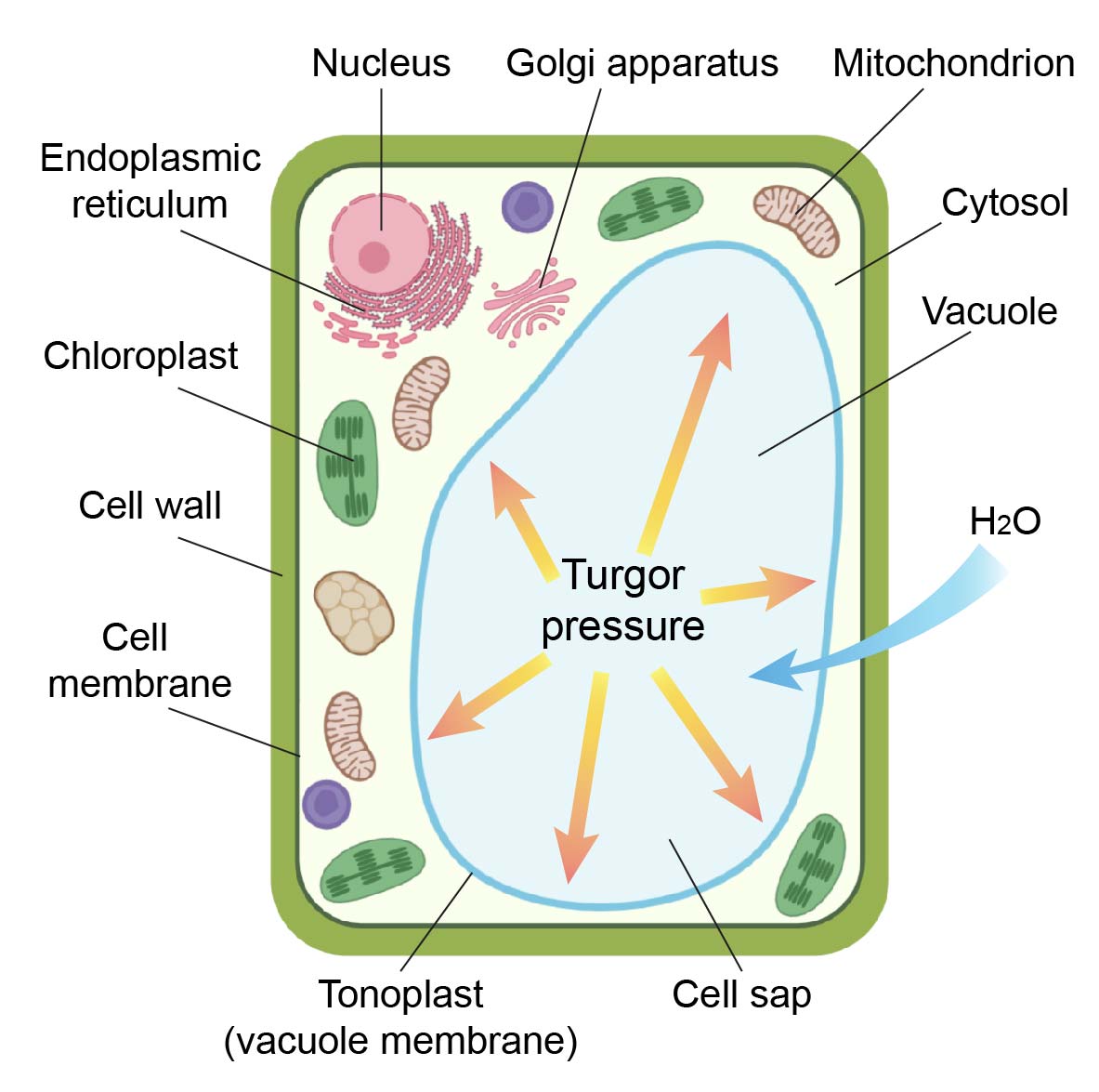
[In this figure] Drawing of a plant cell showing a large vacuole.
Chloroplast
- Chloroplasts are organelles that conduct photosynthesis and produce energy for the plant cells.
- Chloroplasts convert the light energy of the Sun into sugars (a process called “photosynthesis”) that can be used by cells. At the same time, the reaction produces oxygen (O2) and consumes carbon dioxide (CO2).
- Chloroplasts consist of many stacks of sac structures, called thylakoid system. The molecules (Chlorophyll) that absorb the energy of the Sun locate inside the thylakoid sacs.
- Chloroplast plays an important role in plant innate immunity.
- Chloroplasts and mitochondria share many in common. They both have two layers of membranes, their own DNA and ribosomes. They are believed to be derived from endosymbiotic bacteria engulfed by the early ancestors of today’s eukaryotic cells.
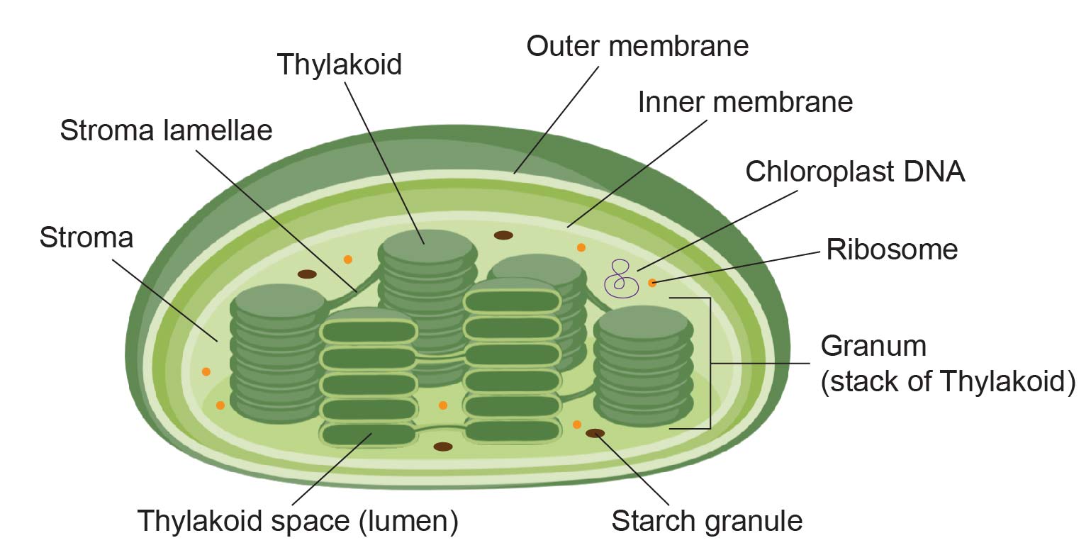
[In this figure] The structure of chloroplast.
Related posts
Animal Cell Model Part I – cell membrane, cytosol, nucleus, and
mitochondria.
Animal Cell Model Part II – endoplasmic reticulum, ribosome, Golgi
apparatus, peroxisome, and lysosomes.
Animal Cell Model Part III – two types of temporary organelles
involving eating behaviors, autophagosomes, and endosomes.
Animal Cell Model Part IV – two types of temporary organelles only
appearing during mitosis, centrosomes, and chromosomes.
Plant Cell Model Part V – cell wall, vacuole, and chloroplast.
More Cell Biology
https://immortalista.blogspot.com/p/cell-biology-cytology_0.html
Applied Cell Biology; watch cells catch the flu bug


